90-y-o Mexican, female
Community Liason
COD: Dementia
Medical History
N/a
Learning Objectives
Exercises on Silent Teacher

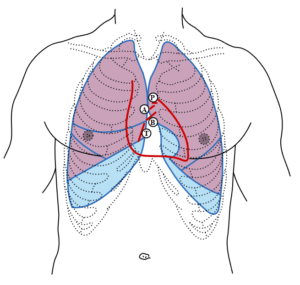
Figure: Front of thorax, showing surface relations of bones, lungs (purple), pleura (blue), and heart (red outline). P. Pulmonary valve. A. Aortic valve. B. Bicuspid valve. T. Tricuspid valve. (Source: Henry Gray (1918) Anatomy of the Human Body. Bartleby.com: Gray’s Anatomy, Plate 1216. Vectorized in Inkscape by Mysid)
Click here to see a 3D artistic model for surface projections of the thorax.
Diaphragm: The diaphragm is a dome-shaped musculofibrous septum separating the abdomen and thorax. It is bounded above by both pleural spaces and the pericardium, which is attached to the central tendon. Structures immediately adjacent to the inferior side of the diaphragm include the liver, spleen, stomach, and to varying degrees the colon, omentum, and small bowel. The origin of the diaphragm includes the lower sternum, lower six costal cartilages, and adjacent ribs, and medial and lateral lumbocostal arches. The crura, two tendinous pillars, arise from the lumbar vertebrae. The insertion of the diaphragm is the central tendon, an aponeurosis, located at the top of the dome, is oriented transversely and separated into three segments. At rest, the diaphragm rises to the level of the fourth intercostal space on the right and the fifth intercostal space on the left.
Liver
The upper limit of the right lobe of the liver, in the middle line, is at the level of the junction between the body of the sternum and the xiphoid process; on the right side, the line must be carried upward as far as the fifth costal cartilage in the mammary line, and then downward to reach the seventh rib at the side of the thorax. The upper limit of the left lobe can be defined by continuing this line downward and to the left to the sixth costal cartilage, 5 cm. from the middle line. The lower limit can be indicated by a line drawn 1 cm below the lower margin of the thorax on the right side as far as the ninth costal cartilage, thence obliquely upward to the eighth left costal cartilage, crossing the middle line just above the transpyloric plane and finally, with a slight left convexity, to the end of the line indicating the upper limit.
Take three points:
(a) 1.25 cm below the right nipple
(b) 1.25 cm below the tip of the tenth rib
(c) 2.5 cm below the left nipple
Join (a) and (c) by a line slightly convex upward; (a) and (b) by a line slightly convex lateralward; and (b) and (c) by a line slightly convex downward.
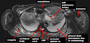
Exercises on silent teachers’ MRI scans
(Please note rad3d requires sign-in/login) (Please watch the video here to learn how to adjust images)
OSTEOLOGY
MUSCLES
VISCERA AND COMPONENTS
SPINAL CORD
Here are some examples


Self Assessment
95-y-o Chinese, female
Farmer
COD: Cerebrovascular disease
Medical History
Stroke, near-term memory loss, 2 falls-injuries to tailbone and shoulder.
Learning Objectives
Exercises on Silent Teacher

Figure: Front of thorax, showing surface relations of bones, lungs (purple), pleura (blue), and heart (red outline). P. Pulmonary valve. A. Aortic valve. B. Bicuspid valve. T. Tricuspid valve. (Source: Henry Gray (1918) Anatomy of the Human Body. Bartleby.com: Gray’s Anatomy, Plate 1216. Vectorized in Inkscape by Mysid)
Click here to see a 3D artistic model for surface projections of the thorax.
Diaphragm: The diaphragm is a dome-shaped musculofibrous septum separating the abdomen and thorax. It is bounded above by both pleural spaces and the pericardium, which is attached to the central tendon. Structures immediately adjacent to the inferior side of the diaphragm include the liver, spleen, stomach, and to varying degrees the colon, omentum, and small bowel. The origin of the diaphragm includes the lower sternum, lower six costal cartilages, and adjacent ribs, and medial and lateral lumbocostal arches. The crura, two tendinous pillars, arise from the lumbar vertebrae. The insertion of the diaphragm is the central tendon, an aponeurosis, located at the top of the dome, is oriented transversely and separated into three segments. At rest, the diaphragm rises to the level of the fourth intercostal space on the right and the fifth intercostal space on the left.
Liver
The upper limit of the right lobe of the liver, in the middle line, is at the level of the junction between the body of the sternum and the xiphoid process; on the right side, the line must be carried upward as far as the fifth costal cartilage in the mammary line, and then downward to reach the seventh rib at the side of the thorax. The upper limit of the left lobe can be defined by continuing this line downward and to the left to the sixth costal cartilage, 5 cm. from the middle line. The lower limit can be indicated by a line drawn 1 cm below the lower margin of the thorax on the right side as far as the ninth costal cartilage, thence obliquely upward to the eighth left costal cartilage, crossing the middle line just above the transpyloric plane and finally, with a slight left convexity, to the end of the line indicating the upper limit.
Take three points:
(a) 1.25 cm below the right nipple
(b) 1.25 cm below the tip of the tenth rib
(c) 2.5 cm below the left nipple
Join (a) and (c) by a line slightly convex upward; (a) and (b) by a line slightly convex lateralward; and (b) and (c) by a line slightly convex downward.
Exercises on silent teachers’ MRI scans
(Please note rad3d requires sign-in/login) (Please watch the video here to learn how to adjust images)
OSTEOLOGY
MUSCLES
VISCERA AND COMPONENTS
SPINAL CORD
Here are some examples


Self Assessment