
Learning Outcomes
By the end of this case, students will be able to:
- Identify gross internal anatomical structures and classify them according to organ system.
- List the main organs of the digestive system
- Locate and identify organs of the digestive system/alimentary canal on an image
- Name and identify the accessory digestive organs, listing a function for each
- Identify the hepatic duct, cystic duct, gallbladder, common bile duct, sphincter of the hepatopancreatic ampulla (ampulla of Vater and sphincter of Oddi) and discuss the roles of those structures in the flow of bile
- Use a stepped approach to critically evaluate the patient medical complaint
- Identify key clinical signs and symptoms to determine a diagnosis
Subject
– 85-year-old Japanese female
– Obesity ( Height 5’2″, weight 220lbs)
– Xanthomas around her right eye
History & Chief Complaint
– Chief Complaint: Abdominal pain and jaundice
– She comes to the ED with 1 to 2 days of slight abdominal pain as well as non-specific pain in the shoulder and neck area. She has increasing nausea and she has vomited. The patient says that the pain started suddenly and continued for several hours. She also presents with yellow discoloration of her eyes which she noticed two days ago while washing her face.
Exam
– The patient was febrile
– Abdominal examination revealed no rebound tenderness
– Skin was icteric
– HDL:34mg/dL (normal range>40 mg/dL ) LDL: 208mg/dL (normal range <130 mg/dL ) Triglyceride:362mg/dL (normal range <150 mg/dL )
– Abnormal liver function tests were noted SGOT 713 IU/L (normal range 5–40 IU/L), SGPT 429 IU/L (3–35 IU/L), alkaline phosphatase 192 IU/L (35–104 IU/L), and total bilirubin 2.8 mg/dl (0.2–1.2 mg/dl).
Radiology Findings
– Biliary stones, sludge, and biliary polyp (Lead to choledocholithiasis and acute cholangitis)
– See the segmented model here.
Differential Diagnosis of Cholelithiasis
– Hepatitis
– Hepatic abscess
– Pancreatitis
– Kidney diseases
– Appendicitis
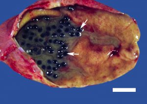
Pathology Findings
– The two major types of gallstones include cholesterol stones and pigment (bilirubin-containing) stones. The pathology image demonstrates many black stones within the gallbladder lumen associated with purulent material, likely reflecting acute cholecystitis. The black color is consistent with pigment (bilirubin-containing) stones.
Anatomical Considerations
– Referred pain can be defined as the convergence of visceral and somatic nerve input. Choledocholithiasis causes gall bladder inflammation that impinges on the mesothelium of the diaphragm causing phrenic sensory nerve fiber stimulation through segments C3,4,5 which converge with somatic sensory input conducted by the supraclavicular nerves of the cervical plexus via C3,4 resulting in referred pain to the shoulder. View the dissection here. Review the cervical plexus and phrenic nerve here.