Parapelvic cysts
Definition
The kidney is prone to cystic lesions with an incidence of about 25% in adults older than 40 years increasing to 66% when the adult is older than 80 years. A parapelvic cyst occurs near the renal pelvis or pedicle. A parapelvic cyst is a non-genetic cyst with pathological changes. The mechanism and tissue structure of para-pelvic cysts are similar to a simple kidney cyst. The cause of this disease in the elderly is acquired renal obstruction while in young people it is generally the result of congenital dysplasia.
Radiology
The question is whether the renal pelvis and calyces are dilated or stretched since parapelvic cysts do not communicate with the collecting system and are probably lymphatic in origin or develop from embryonic remnants Parapelvic cyst can mimic hydronephrosis and so can be confused with pelvi-ureteric junction obstruction as the ureter in both conditions is not dilated. This could be well differentiated by CT urography and is likely an incidental finding on an MR.
Anatomical Correlations & Exercises
Examine the internal features of the kidney. A simple renal cyst is a benign condition usually discovered as an incidental finding. However, on occasions, complications within the cyst occur to produce unusual symptoms, which make diagnosis difficult. Most cysts are placed superficially and expand away from the kidney and can, on occasions, reach sufficient size to press on neighboring structures, such as the stomach, to produce dyspepsia. Complications that have been reported, include rupture either spontaneous or from trauma, infection, haematuria, rarely hypertension.
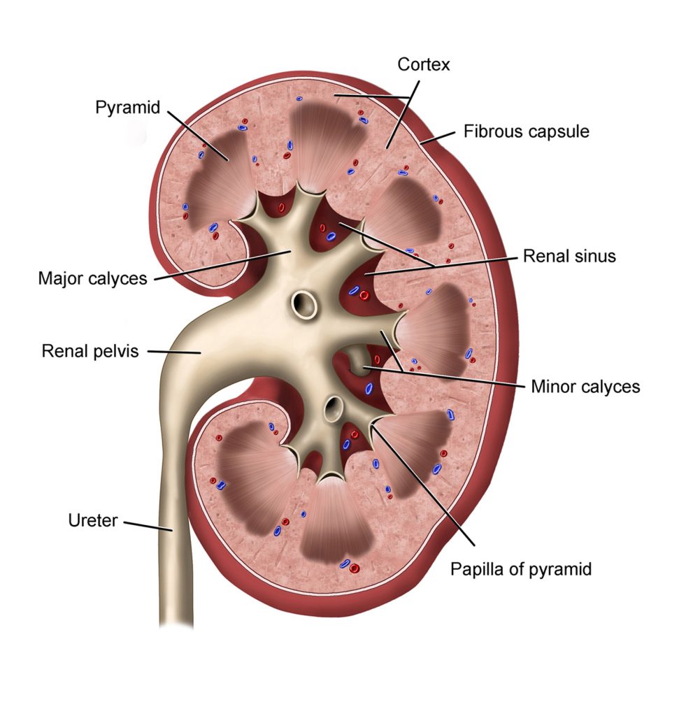
The kidney receives the renal artery and vein which enter the substance of the kidney through its hilum. The renal artery is a tributary of the abdominal aorta while the renal vein drains into the inferior vena cava. The renal pelvis lies posterior to these renal vessels and the ureter is the inferior continuation of the renal pelvis. On hemisection, numerous structures can be seen in the kidney. A fibrous capsule envelopes the kidney. A renal cortex forms the outer 1/3 of the kidney which will provide as renal columns into the inner kidney core or renal medulla. Renal pyramids consist of collecting ducts and lie between the renal columns. The filtrate from renal pyramids travels through a renal papilla and into a minor calyx. Approximately 3-4 minor calyces empty into a major calyx. The waste eventually exits the kidney through the renal pelvis and ureter.
Using the picture below, identify the internal structures of the kidney.

